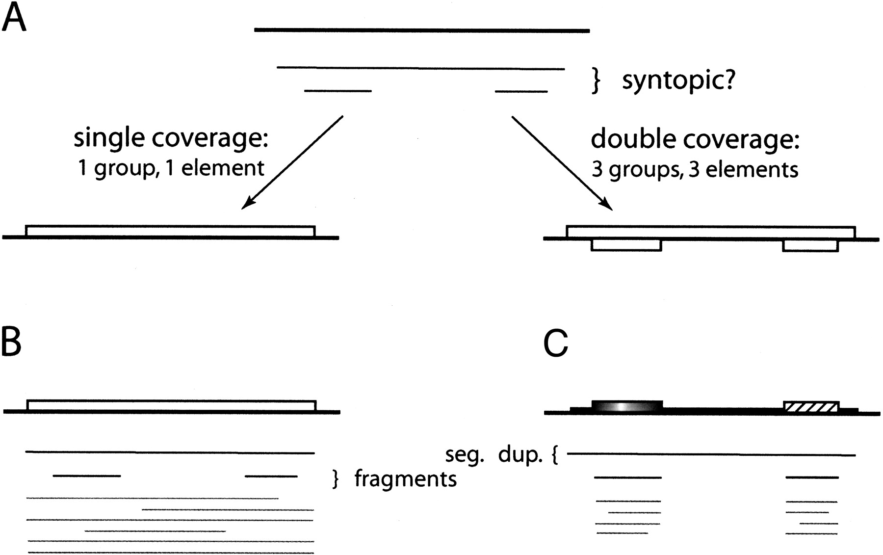
Figure 2.
Different biological scenarios require different methods of syntopy inference. (A) For three images (thin black lines) in a genomic region (top bold black line), the single and double coverage methods lead to different definitions of elements. (B) A full-length element and its images (black and grey lines below). The top long image is formed with another full-length member in its family, whereas the shorter images are formed with the fragmented members. (C) A segmental duplication covering two kinds of elements. The top long image is formed with the other copy of this segmental duplication, whereas the shorter images are formed with other members in the two families, respectively.