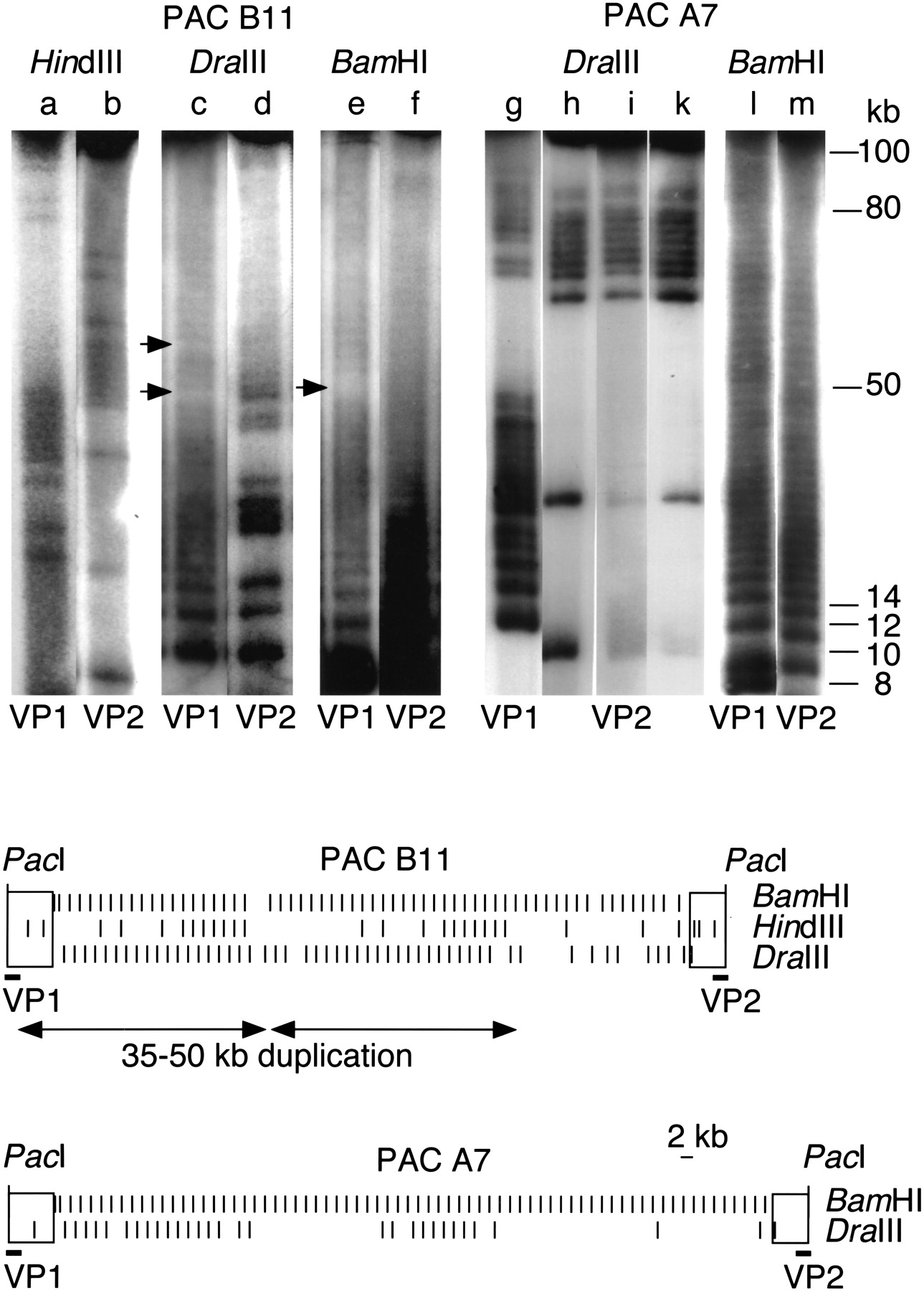
(Top) Large-scale partial restriction mapping. Intact PAC DNA was linearized using the unique PacI site in vector DNA, and electroeluted (Biotrap, Schleicher Schüll). Partial digestions, using 0.1–3 U and 1–2 min of incubation time, were run on a CHEF DRII pulsed-field gel apparatus (Biorad) under conditions separating fragments up to 80–100 kb (switch 2.8 sec, 6 V/cm, 25 h). Gels were Southern-blotted and hybridized with end probes VP1 and VP2. X-Ray images were photographed and processed on a Nicon Cool Scan III. Each lane (a–m) represents one selected condition. PAC B11 (lanes a–f) shows clustered occurrence of the HindIII variant common within this array segment. Two clusters of 7 higher-order repeats containing the HindIII variant are arranged in tandem, at a distance of ∼50 kb (lanesa,b). The slightly more diverged array of PAC B11 shows disruption of the 2-kb BamHI higher-order repeat structure (lane e) and of the DraIII variant (lanec), possibly marking sites of unequal recombination (little arrows). The highly homogeneous PAC A7 (lanes g–m) is entirely composed of 2-kb BamHI higher-order repeats (lanesl,m). The DraIII variant, which is presently in a fixed transition state, occurs in clusters (lanesg–k), indicating a rather localized component of spread. Two additional, independent colonies were consecutively replated for 6 d (400 generations), which did not alter theDraIII restriction patterns (lanes i,k), further demonstrating the high stability of α-satellite DNA cloned in PACs. Integrated restriction maps of PACs B11 and A7 are shown at the bottom. Left (VP1) and right (VP2) end probes (bars) are indicated below PAC vectors (open rectangles). Variants presently in transition (HindIII, DraIII) occur in a highly nonrandom fashion, forming clusters of varying size. PAC B11 presents a duplicated pattern of 35–50 kb (duplicate arrows).