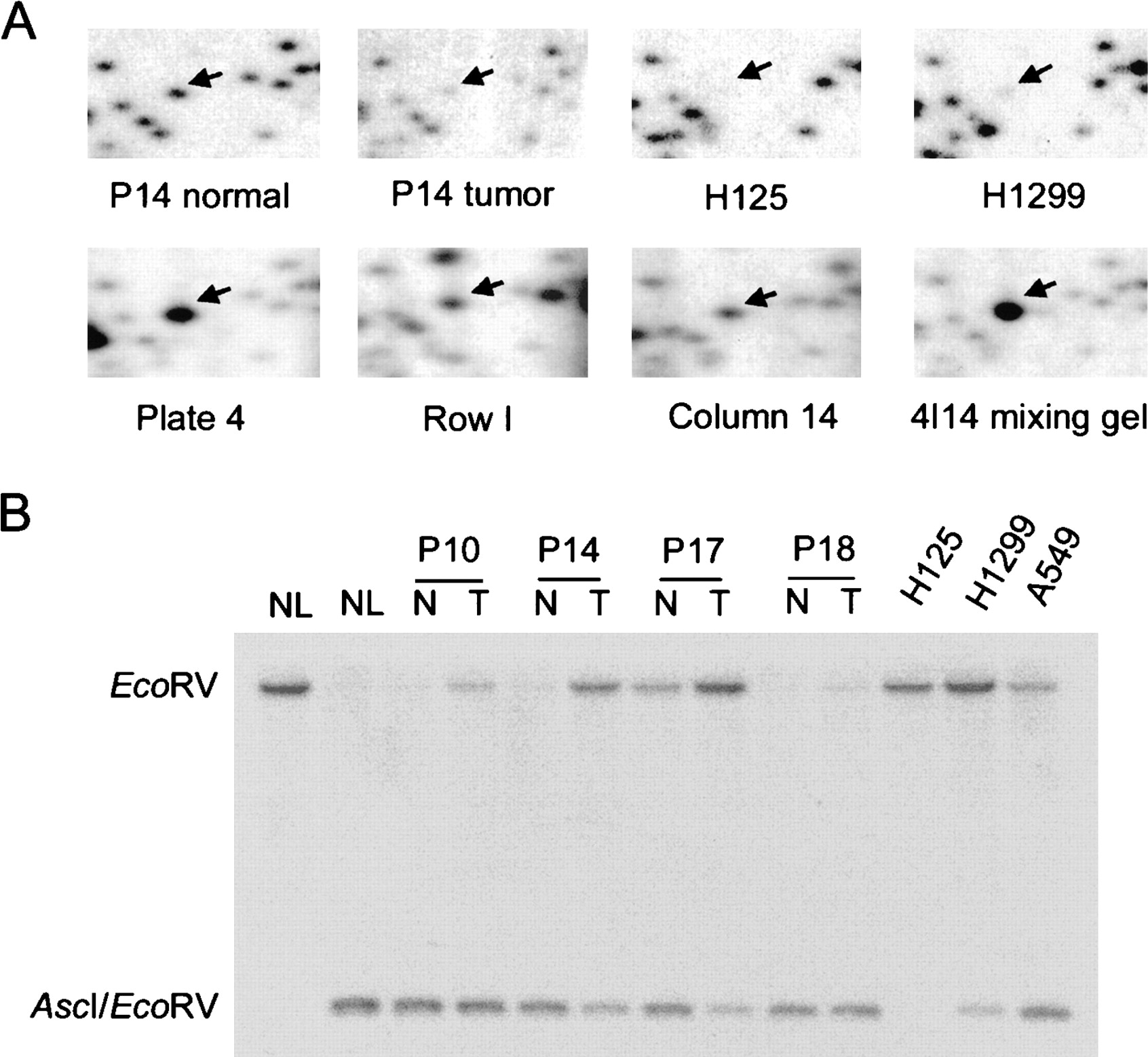
RLGS identifies DNA methylation in primary lung cancer. (A) Sections from RLGS profiles including RLGS fragment A2E54 (arrow). Sections from normal and tumor profiles from patient 14 as well as two lung cancer cell lines (H1299 and H125) are shown. The correspondingAscI–EcoRV clone was found in plate 4, row I, and column 14, and this clone was confirmed by use in a mixing gel. (B) DNA from AscI clone 4I14 corresponding to RLGS spot A2E54 was used for Southern analysis. DNAs from normal lung (NL), lung tumors (T), and adjacent normal tissue (N) from patients 10, 14, 17, and 18, as well as from three lung cancer cell lines H125, H1299, and A549 were digested with AscI and EcoRV. DNA in the first lane was digested only with EcoRV and shows the size of the EcoRV fragment. In the double digests, hybridization to the large EcoRV band is indicative of protection of theAscI site digestion by methylation. The smaller band is indicative of cutting by AscI.