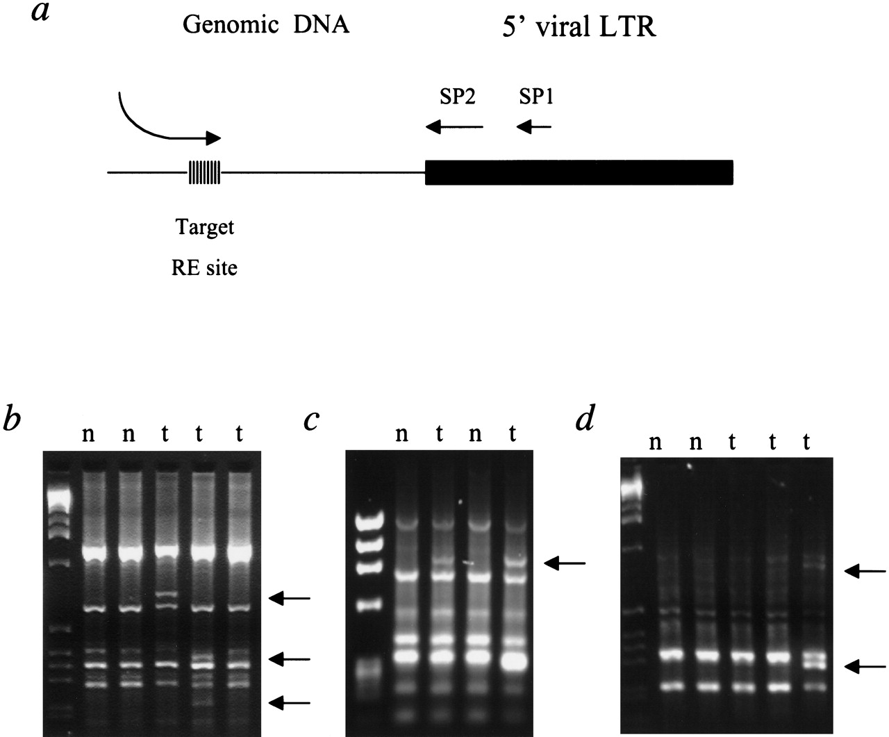
VISA PCR using MuLV LTR-specific primers. (a) Schematic representation of VISA. Viral insertion sites are amplified using primers specific for the long terminal repeat (LTR) of the MuLV in combination with RE-anchored degenerate primers that amplify from flanking genomic DNA. Two rounds of PCR are performed on unmodified genomic DNA using nested virus-specific primers to ensure specificity. Successful amplification of any viral insertion site is dependent on whether the RE anchor sequence is present in the genomic sequence flanking the viral LTR. Therefore, to increase the number of insertion mutations amplified, a minimum of five RE-anchored degenerate primers are used on each sample. The approximate binding sites for LTR specific primers SP1 and SP2 are shown. The RE-anchored degenerate primer contains an M13F linker (shown as a curved line) that is not expected to bind mouse DNA during the first round of PCR, but is useful for subsequent amplifications and sequencing. The stripped box indicates the location of the restriction enzyme recognition site target for the RE-anchored degenerate primer. (b–d) Representative examples of VISA products generated with RE-anchored primers specific for HindIII (b), EcoRI (c), andBclI (d). DNA isolated from brain tissue, which is rarely infiltrated by tumor cells, was used as a control to identify all nontarget amplification products such as endogenous proviral insertion sites, internal provirus sequences, or single primer amplification products generated by the restriction-site anchored primer. Tumor-specific VISA products, ranging in size from 50 bp to 2.0 Kb were gel purified and sequenced directly (identified by arrows). Lane designations are (n) (nontumor); (t) tumor.